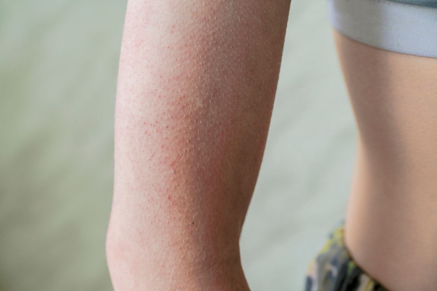
Wyprysk z hiperkeratozą - Objawy, Diagnoza i Leczenie
ROGOWIEC
Choroba genetyczna cechująca się hyperkeratynizacją skóry i paznokci. Jest to pogrubienie ograniczone do powierzchni dłoniowych i podeszwowych, pojawiające się zwykle z powodu mutacji.
GŁÓWNE OBJAWY ROGOWCA:
- nadmiernie zrogowaciały naskórek
- żółte lub woskowe zabarwienie skóry
- zgrubiałe i przerosłe płytki paznokciowe
Odmiany rogowca
Unna-Thost pojawia się ok. 1-2 roku życia. Zmiany rogowe są symetryczne, występuje nadpotliwość dłoni i stóp.
Keratoma disseminatum pojawia się po 20 roku życia. Charakteryzuje się drobnymi, rozsianymi wykwitami. Z wiekiem może ich przybywać.
Keratoma trnsgrediens et progrediens pojawia się w pierwszych miesiącach życia. Ogniska hyperkeratotyczne występują poza dłońmi i stopami i najczęściej znajduję się na łokciach i kolanach.
Lokalizacje odcisku:
- palce stóp (okolica grzbietowa stawy międzypaliczkowe, boczna i przyśrodkowa powierzchnia palców w miejscu stykania się skóry Clavus mollis, okolica podeszwowa palca – Clavus appex)
- przodostopie (głowy kości śródstopia)
- wały paznokciowe (clavus sulcus)
- przestrzenie podpaznokciowe (clavus subungualis)
Clavus durus (Cd) – zbudowany jest ze zwartej i twardej masy ułożonej warstwowo (nawet do 200 warstw), zawierający jądro.
Clavus mollis (Cm) – to inaczej odcisk miekki.
Clavus vascularis (Cv) – to odcisk z zawartością drobnych naczyń krwionośnych.
Clavus neurovascularis (Cnv) – odcisk nerwowo – naczyniowy.
Clavus neurofibrosis (Cnf) – odcisk nerwowo – włóknisty.
Clavus papilaris (Cp) – to odcisk brodawkowy. Często się powtarza.
Clavi miliares (Cmil) – odciski mnogie.
Lokalizacje modzeli:
- palce stóp (okolica grzbietowa i podeszwowa oraz boczna)
- pięta
- przodostopie
- boczna krawędź stopy
ODCISK
BRODAWKA WIRUSOWA
Często występują u dzieci
Dr n.med. Danuta Nowicka ”Dermatologia. Ilustrowany podręcznik dla kosmetologów”, Wrocław 2014
HYPERKERATOZY – rodzaje i różnicowanie cz.2
- odciski
- modzele
- nadmierne rogowacenie i zespół pękających pięt
- rogowiec
Zmiany będące reakcją obronną skóry na bodźce zewnętrzne. Powstają na skutek nadmiernego wytwarzania komórek warstwy rogowej naskórka. Powstaje bariera uniemożliwiająca fizjologiczną migrację nowych komórek, a co za tym idzie dochodzi do pozostawania korneocytów w niższych warstwach skóry.
Odciski są wyniosłymi zgrubieniami naskórka o kształcie okrągłym, podłużnym lub nieregularnym. Cechą wyróżniającą odciski jest obecność rdzenia, czyli twardego czopu rogowego, zlokalizowanego najczęściej centralnie.
Trzpień odcisku często sięgając głęboko (nawet do okostnej), drażni zakończenia nerwowe dając uczucie bólu.

 U nas zapłacisz kartą
