 U nas zapłacisz kartą

Rak podstawnokomórkowy - Prognozy i leczenie
Basal cell carcinoma is the most common type of cancer
If you’ve been diagnosed with basal cell carcinoma (BCC), you have plenty of company. Basal cell carcinoma is the most common type of skin cancer. It’s also the most common type of cancer. Doctors diagnose millions of people with basal cell carcinoma every year.
You have a greater risk of developing this skin cancer if you have a lighter skin tone and seldom protected your skin from the sun throughout your life or used tanning beds.
People of all skin tones develop basal cell carcinoma. However, people who have light skin that rarely tans and tends to freckle, red or blond hair, and light-colored eyes have a greater risk of developing this skin cancer.
Before basal cell carcinoma develops, people with lighter skin tones often notice signs of sun damage on their skin, such as age spots, patches of discolored skin, and deep wrinkles. These signs can develop years before cancer.
Treatment / Management
event recurrence at a later date, (2) to correct any functional impairment resulting from the tumor, and (3) to give the best cosmetic result to the patient, especially because most BCCs are on the face.[11]
Treatment of BCC is usually surgical, but some forms of BCC are amenable to medical treatment or radiation therapy. The various types of therapy include Mohs micrographic surgery (MMS), standard surgical excision, EDC, radiation, photodynamic therapy, cryosurgery, topical therapies, and systemic medications such as Vismodegib. The recurrence rates for primary BCC are as follows: Mohs surgery, 1.0%, surgical excision, 10.1%, EDC, 7.7%, radiation therapy, 8.7%, and cryosurgery, 7.5%.
Mohs surgery provides the best long-term cure rate of any treatment modality for BCC. MMS is the gold standard for treating high-risk BCCs and recurrent BCCs because of its high cure rate and tissue-sparing benefit. The high cure rate is attributed to an examination of 100% of all the tissue margins when compared with standard vertical sectioning, wich only examines less than 1% of the outer peripheral and deep margins. By only taking thin tissue layers from the areas with positive tumor margins, the wound size is minimized, and a superior cosmetic outcome can be expected.
Radiation therapy is a primary option for treating BCC or SCC if surgery is contraindicated. It also can be used as an adjuvant treatment for basal cell carcinoma when further surgery could sacrifice major nerves or other vital structures, or there is a perineural invasion by cancer cells. The disadvantages of radiation therapy are cost, poor cosmesis in some patients, prolonged course of treatment (15 to 30 visits), and increased risk for future skin cancers. Scars from radiation therapy tend to worsen with time, while surgical scars improve over time.
Topical therapy is another treatment for basal cell carcinoma. Topical 5-fluorouracil (5-FU) and Imiquimod 5% cream are approved by the Food and Drug Administration (FDA) to treat superficial BCC. Both topical therapies are good options in patients with multiple superficial BCCs and in patients who are poor surgical candidates. Application site reactions are common and include erythema, pruritus, pain, edema, hypopigmentation, hyperpigmentation, crusting, bleeding, and erosions. Another disadvantage is there no histologic confirmation of complete tumor clearance.
Rak kolczystokomórkowy (carcinoma spinocellulare)
Rak kolczystokomórkowy (carcinoma spinocellulare) jest nowotworem znacznie rzadziej występującym niż rak podstawnokomórkowy. Rak kolczystokomórkowy najczęściej pojawia się u osób w wieku średnim i starszym. Cechuje się szybkim wzrostem i dużą złośliwością, wykazując skłonność do naciekania podłoża (czyli wrastania komórek raka do tkanek położonych pod guzem) i szerzenia się drogą przerzutów. Punktem wyjścia mogą być stany przedrakowe, zwłaszcza rogowacenie słoneczne i róg skórny. Zmiany zlokalizowane są najczęściej na granicy błon śluzowych i skóry (czerwień wargowa – szczególnie wargi dolnej), w okolicach nosa, oczodołów, narządów płciowych. Rak może też lokalizować się na powiekach. Przeczytaj więcej: Rak kolczystokomórkowy powiek
Do objawów niepokojących należy zaliczyć: gwałtowny wzrost masy guza, stwardnienie, tworzenie się owrzodzeń oraz krwawienia. Przebieg raka kolczystokomórkowego jest zależny od umiejscowienia, rozległości i stopnia naciekania podłoża. Skłonność do przerzutów jest znacznie mniejsza w przypadku zmian wywodzących się z rogowacenia słonecznego, a zwiększa się w przypadku rozwoju nowotworu na podłożu owrzodzeń, blizn i przewlekłych stanów zapalnych.
Rak brodawkujący (carcinoma verrucosum)
Rodzajem raka kolczystokomórkowego jest rak brodawkujący (carcinoma verrucosum). To nowotwór występujący na narządach płciowych, w jamie ustnej i w obrębie stóp. W raku brodawkującym stwierdzono wirusy brodawczaka ludzkiego HPV 6 i HPV 11. W przypadku raka brodawkującego umiejscowionego w jamie ustnej istotnym czynnikiem ryzyka jest używanie tytoniu (żucie, wciąganie tabaki do nosa), natomiast w przypadku raka zlokalizowanego na powierzchni podeszwy stopy czynnikiem takim są urazy. Wzrost raka brodawkującego jest bardzo powolny, jego powierzchnia jest pokryta masami rogowymi. Na ogół nie daje przerzutów.
Is basal cell carcinoma serious?
For most people, basal cell carcinoma is not life-threatening. This skin cancer tends to grow slowly. It seldom spreads to another part of the body. Even so, treatment is important.
Over time, basal cell carcinoma can grow wide and deep. It can spread deeply into the skin, wrap around nerves and blood vessels, and invade muscles and bone. When the cancer grows deep, it can change the way you look. For some people, this can be disfiguring.
When found early, this skin cancer is highly treatable. An early basal cell carcinoma can often be removed during an appointment with your dermatologist.
One common sign is a slowly growing, non-healing spot that sometimes bleeds. Basal cell carcinoma can also appear on the skin in other ways.
You’ll find the signs and symptoms along with several pictures of this skin cancer at, Basal cell carcinoma: Signs and symptoms.
Image
Getty Image
References
Cameron MC, Lee E, et al. “Basal cell carcinoma: Epidemiology, pathophysiology, clinical and histological subtypes, and disease associations.” J Am Acad Dermatol 2019,80:303-17.
Gloster HM, Neal K. “Skin cancer in skin of color.” J Am Acad Dermatol 2006,55:741-60.
Nouri K, Ballard CJ, et al. “Basal cell carcinoma.” In: Nouri K, et al. Skin Cancer. McGraw Hill Medical, China, 2008: 61-81.
Written by:
Paula Ludmann, MS
Reviewed by:
Carrie L. Kovarik, MD, FAAD
Natalie H. Matthews, MD, FAAD
Darrell S. Rigel, MD, FAAD
Last updated: 4/28/23
Reproduction or republication strictly prohibited
without prior written permission.
History and Physical
Many clinical variants of BCC exist, but the most recognized types are superficial, nodular, and morphea-like BCC. Nodular BCC is the most common (see Image. Nodulocystic Basal Cell Carcinoma).
A skin biopsy is necessary for clinical confirmation of BCC. A shave, punch, or excisional biopsy are all options, taking care to include some portion of the dermis in the specimen to differentiate between superficial and other invasive histologic subtypes of BCC. It should be noted that punch and shave biopsy techniques are about 80% accurate in diagnosing the various subtypes of basal cell carcinoma.[10]
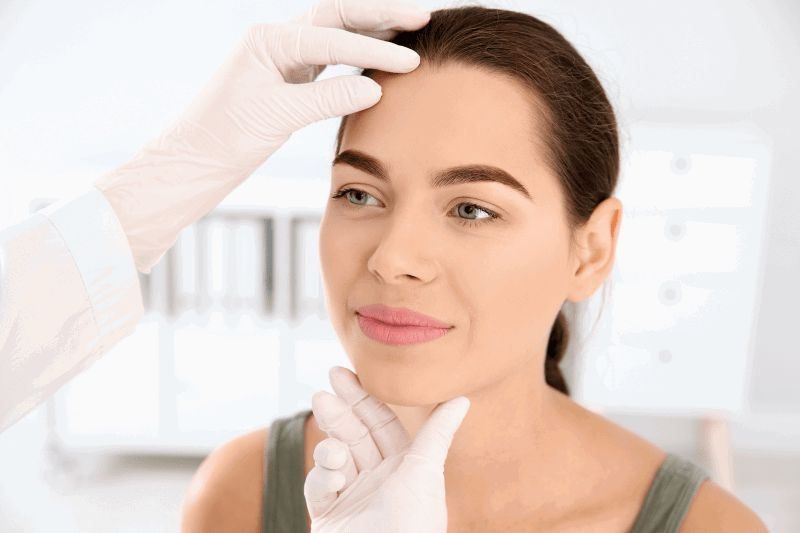
A qualified practitioner should perform a complete skin examination since individuals with one finding of skin cancer often have additional cancers or pre-cancers at other sites and have an increased risk of developing malignant melanoma. Documentation of the location of the lesions with photographs or digital images is a recommended procedure. There is a low threshold for obtaining skin biopsies in these patients. Preoperative imaging studies may be necessary when there is suspicion of parotid gland, muscle, deep soft tissue, orbital, bone involvement, or perineural invasion. Patients with a history of BCC should have long-term, even lifetime, follow-up, particularly those with multiple or high-risk tumors.
Dermoscopy can be beneficial to the experienced clinician, aiding in the diagnosis of non-pigmented and pigmented BCCs. The hallmark of BCC on dermoscopy is the presence of well-focused arborizing vessels. Additional findings include multiple blue-gray globules, leaf-like structures, large blue-gray ovoid nests, and spoke-wheel areas. There is no pigment network, as one would see with dermoscopy of pigmented lesions.